
DLD Home> FAQs & Forms
Sample Submission – Frequently Asked Questions
> Ensure maximum 48 hours transit time during the hottest months.
> Do not ship before holidays / long weekends.
> Proper packaging is essential for safety and compliance – follow all regulations for labeling and transport of diagnostic specimens. Reference Fedex Guide: https://www.fedex.com/content/dam/fedex/us-united-states/services/UN3373_fxcom.pdf.
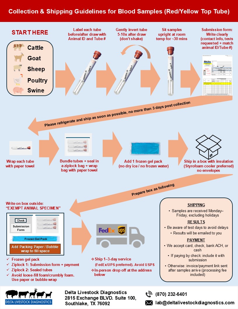
> Each tube must be clearly labeled with both the tube number and animal ID, which must correspond to the information provided on the submission form. Submission forms must be completed legibly.
> We recommend not using the United States Postal Service (USPS) for diagnostic shipments. Even if shipped express, USPS deliveries often take an additional 2 days. For faster turnaround, use FedEx or UPS.
> Primary Container: Use a watertight, leakproof container (e.g., 10 mL ear notch tube, red/yellow top tube, or screw-cap tube). Label each sample with animal ID.
> Secondary Packaging: Wrap each primary container in absorbent material (e.g., paper towel) and place in a leakproof zip-seal bag. Separate multiple tubes in a single ziplock bag with absorbent material. Wrap ziplock bag with a paper towel.
> Outer Packaging: Use sturdy cardboard box (pregnancy samples) or insulated cooler (disease samples). Include ice packs (not frozen water bags) that are seperated from the secondary pacakaging by paper towels or bubble wrap. Must withstand a 4-foot drop test.
> Submission Form and any Payment: Place in waterproof bag. Ensure contact details (address, phone, email) are included on the submission form.
> Labeling: Mark as “Exempt Animal Specimen” on the outer packaging.
> Do not send samples in syringes with needles attached (packages with sharps will be rejected).
> Do not use gloves, OB sleeves, or leaky containers for sample transport.
> Protect all samples from heat and freezing — hemolyzed or frozen samples may be rejected.
> Collect whole blood into red-top / yellow-top tubes (no anticoagulant).
> Gently invert tube 5-10 times after drawing the sample (don’t shake).
> Allow tubes to clot upright at room temperature for 30 minutes before refrigeration.
> After clotting, refrigerate at 2–8 °C until shipping.
> Do not freeze.
(Red/yellow top tubes for tests such as Johne’s ELISA, CAE/OPP, CL)
> Collect whole blood into red-top / yellow-top tubes (no anticoagulant).
> Gently invert tube 5-10 times after drawing the sample (don’t shake).
> Allow tubes to clot upright at room temperature for 30 minutes before refrigeration.
> After clotting, refrigerate at 2–8 °C until shipping.
> Do not freeze.
> Collect whole blood into purple-top tube (EDTA anticoagulant).
> Invert gently to mix — do not shake.
> Keep refrigerated (2–8 °C) until shipping.
> Do not freeze.
> Collect tissue in a 10 mL ear notch tube (preferred) or other sterile, leakproof tube.
> Keep refrigerated (2–8 °C) until shipping.
> Do not freeze.
> Must be fresh and collected directly from rectum (not ground).
> Submit in 10 mL ear notch tube (preferred) or other leakproof sterile hard plastic container.
> Avoid overfilling to prevent leakage.
> Place the leakproof container into a ziploc bag.
> Do not freeze.
> Ship overnight to lab as soon as possible to prevent degradation and egg hatching.
> Collect pus from abscess using sterile syringe (no needle / syringe for shipping) or swab.
> For swabs, use sterile polyester (Dacron) or nylon flocked swabs with plastic shafts. Avoid cotton, calcium alginate, or wooden shaft swabs.
> Place in 10 mL ear notch tube (preferred) or other sterile, leakproof screw-cap tube.
> Ship overnight to lab to prevent nucleic acid degradation.
> Collect into 10 mL ear notch tube (preferred) or other sterile, leakproof tube.
> Avoid manure/debris contamination.
> Keep refrigerated until ready to ship (2–8 °C).
> Collect aseptically from vesicles using sterile syringe (no needle for shipping) or swab.
> Place in 10 mL ear notch tube (preferred) or other sterile, leakproof tube.
> Keep refrigerated until ready to ship (2–8 °C).
> Ship overnight on ice packs.
> Collect using rope-chew method or sterile swab.
> Transfer into 10 mL ear notch tube (preferred) or other sterile, leakproof tube.
> Use sterile polyester (Dacron) or nylon flocked swabs with plastic shafts. Avoid cotton, calcium alginate, or wooden shaft swabs.
> Keep refrigerated until ready to ship (2–8 °C).
> Ship overnight on ice packs.
> Use sterile polyester (Dacron) or nylon flocked swabs with plastic shafts. Avoid cotton, calcium alginate, or wooden shaft swabs.
> Nasal Swab: Collect from both nostrils with sterile swab.
> Place each swab into an individually labeled sterile, leak-proof tube (e.g., ear notch collection tube) containing approximately 0.5 mL of sterile PBS, sterile 0.9% saline, or VTM.
> If not available, swabs may be submitted dry (reduced sensitivity).
> Keep refrigerated until ready to ship (2–8 °C).
> Ship overnight on ice packs.